AACR Journals Editors’ Picks for June
This month, the editors of the AACR’s journals have chosen to highlight an analysis of birth characteristics and risk of early-onset synovial sarcoma, early research aimed at reducing intestinal toxicity brought on by radiotherapy, and a multicenter study evaluating how patients with cancer fare when hospitalized with COVID-19, among other interesting studies. As always, the articles highlighted here are freely available for a limited time.
Journal: Clinical Cancer Research (June 15 issue)
Leptomeningeal carcinomatosis, a type of metastasis where cancer cells spread to the meninges, can occur in many types of cancer, but is observed most frequently in melanoma, lung, gastrointestinal, and breast cancers. Treatment options for leptomeningeal carcinomatosis remain limited, as few therapeutic strategies can effectively penetrate the blood-brain barrier or blood-cerebrospinal fluid barrier. The taxane derivative ANG1005, which is composed of three paclitaxel molecules covalently linked to the oligopeptide Angiopep-2, was designed to cross central nervous system barriers through receptor-mediated transcytosis, and has shown clinical responses in patients with brain metastases of breast cancer. In this phase II clinical trial, ANG1005 was evaluated in 72 patients with recurrent metastases from breast cancer; 28 of these patients had leptomeningeal carcinomatosis. Among the 60 patients evaluable for intracranial response, nine (15 percent) had a partial response and 32 (53 percent) had stable disease. In the leptomeningeal carcinomatosis subset, nearly 80 percent of patients had intracranial disease control. The authors note that a phase III trial comparing ANG1005 with physician’s best choice of treatment in patients with HER2-negative breast cancer with newly diagnosed leptomeningeal disease and previously treated brain metastases is currently underway. This article was featured on the cover and was highlighted in the June 15 issue.
Journal: Molecular Cancer Therapeutics
Modulation of the immune checkpoint protein PD-1 has been a central tenet of immunotherapy treatments. The PD-1 inhibitor pembrolizumab (Keytruda) was first approved by the U.S. Food and Drug Administration (FDA) in 2014 for the treatment of certain patients with melanoma and has received approvals for a variety of indications since that time. Because recent research is focused on improving PD-1-related immunotherapeutic strategies, the authors further characterized pembrolizumab through biophysical analyses and toxicology studies performed in cynomolgus monkeys. Through fluorometric microvolume assay technology (FMAT), the authors demonstrated that pembrolizumab blocks the binding of PD-1 to both of its ligands, PD-L1 and PD-L2. Using nuclear magnetic resonance (NMR) spectroscopy, the authors characterized interactions between PD-1 and pembrolizumab and identified specific PD-1 residues that were affected by pembrolizumab binding. The authors also characterized interactions between the PD-1 inhibitor nivolumab (Opdivo) and PD-1 using NMR and confirmed that the two PD-1 inhibitors have unique binding sites within the PD-1 protein. In preclinical evaluation, the authors found that treatment of human and primate T cells with pembrolizumab enhanced antigen-specific T-cell IFNγ and IL2 production in ex vivo and in vitro studies. When the researchers performed a toxicological evaluation of pembrolizumab in cynomolgus monkeys, they found that the treatment was well-tolerated, with no adverse findings observed during antemortem and terminal evaluations. The authors conclude that the preclinical data for pembrolizumab are consistent with observed findings demonstrated in human clinical trials. This article was highlighted in this issue.
Journal: Cancer Epidemiology, Biomarkers & Prevention
Birth Characteristics and Risk of Early-Onset Synovial Sarcoma
Synovial sarcoma is a rare, but aggressive, soft tissue sarcoma typically found in children and young adults. While the risk of other forms of sarcoma have been found to be associated with parental age, gestational age, birth weight, and birth order, the etiology of synovial sarcoma remains unclear. In this study, the authors reviewed data from California birth records and the California Cancer Registry to understand the factors that might contribute to synovial sarcoma. They analyzed birth characteristic data of 244 patients who were born between 1978 and 2015 and diagnosed with synovial carcinoma between 1988 and 2015 in California; the analysis also included a control group of 12,200 individuals who were born in the same time frame and had not been diagnosed with any cancer before 35 years of age. The authors found that synovial sarcoma was more common in Hispanics than in non-Hispanic whites. Among Hispanics, higher birth weight increased risk, with a 22 percent increase in risk with every 500-gram increase in birth weight. In addition, birth order was found to influence risk across racial and ethnic groups, as the highest risk of the disease was seen in first-born children. The authors conclude that risk of synovial sarcoma is influenced by several nutritional, developmental, and environmental factors. This article is highlighted in this issue.
Journal: Clinical Cancer Research (June 1 issue)
In colorectal cancer, mutations in the BRAF gene can occur under different transcriptional and immunological settings, which could influence the cancer’s response to treatment. Two transcriptional subtypes of the BRAF V600E mutation have been described in colorectal cancer based on the transcriptional and immunological context under which they occur. The BRAF mutation 1 (BM1) subtype occurs in the context of KRAS/AKT pathway activation, epithelial-mesenchymal transition, and increased immune reactivity, while the BM2 subtype occurs in the context of deregulated cell cycle checkpoints. Here, the authors examined how each subtype responded to a treatment regimen consisting of dabrafenib, trametinib, and panitumumab, which are inhibitors of BRAF, MEK, and EGFR, respectively. They found that patients with the BM1 subtype of colorectal cancer had higher response rates, progression-free survival, and overall survival than patients with the BM2 subtype. The authors suggest that BRAF mutation subtype could be used as a predictive biomarker for therapeutic response in colorectal cancer. This article is highlighted in this issue.
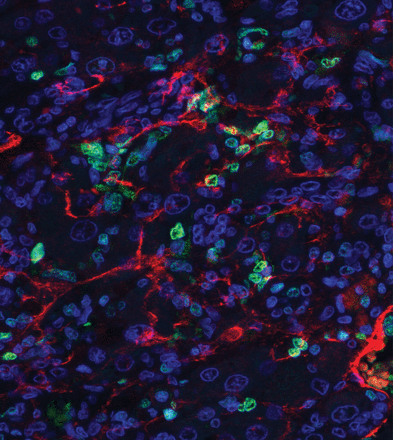
Journal: Cancer Immunology Research
Donor Lymphocyte–Derived Natural Killer Cells Control MHC Class I–Negative Melanoma
The transfer of natural killer cells from a donor to a patient is an effective treatment against certain cancers. Donor lymphocyte infusion (DLI) to deliver natural killer cells after bone marrow transplantation has become an established procedure to treat multiple cancers; however, it is often associated with the occurrence of graft-versus-host disease. Using mouse models of melanoma, the authors of this study demonstrated that DLI without prior bone marrow transplantation elicited infiltration and persistence of the transferred natural killer cells, leading to melanoma control without graft-versus-host disease. Furthermore, DLI was associated with expansion and activation of tumor-infiltrating natural killer cells expressing cytotoxic molecules and maturation signatures. The development of tumor-infiltrating natural killer cells was dependent on IL2 production by CD4+T cells, as IL2 blockade led to impairment of natural killer cell-dependent melanoma control. The authors conclude that DLI without prior bone marrow transplantation can lead to tumor infiltration and expansion of natural killer cells and melanoma control. This study was featured on the cover of this issue.
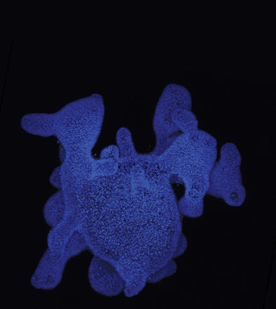
Journal: Cancer Prevention Research
Hepatocellular carcinoma (HCC) develops in chronically inflamed and damaged liver tissue. Previous research has suggested that epigallocatechin gallate (EGCG), an extract from green tea, can suppress liver inflammation and fibrosis in animal models, but its chemopreventive potential remained unclear. In this study, the authors tested the chemopreventive potential of EGCG in a rat model of HCC. Rats were injected with diethylnitrosamine to induce liver cirrhosis and HCC and received EGCG through drinking water. EGCG was well tolerated and was associated with improved serum liver markers and reduced formation of HCC. In addition, rats that received EGCG had decreased expression of genes associated with the Hoshida high-risk gene signature, a signature that is linked to poor prognosis. Furthermore, EGCG inhibited fibrosis progression and induced senescence in hepatic stellate cells. The authors conclude that EGCG exhibited chemopreventive activity in rat models of HCC. This article is featured on the cover of this issue.
Journal: Cancer Research (June 1 issue)
Fibroblast growth factors (FGFs) are elevated in patients with multiple myeloma and correlate with disease activity. However, the mechanism by which FGFs affect disease progression remained unclear. Here, the authors found that FGF and FGF receptor (FGFR) signaling promoted cell survival and disease progression by protecting multiple myeloma cells from oxidative stress-induced apoptosis in multiple myeloma cell lines. Inhibition of FGF/FGFR signaling induced mitochondrial oxidative stress, DNA damage, and apoptosis, thereby impairing the growth and migration of multiple myeloma cells in culture and in animal models. In addition, the authors found that the mitochondrial oxidative stress induced by FGF/FGFR inhibition was due to degradation of the c-MYC oncoprotein and subsequent depletion of glutathione. The authors confirmed these findings in primary multiple myeloma cells cultured from patients with newly diagnosed or relapsed/refractory disease. Together, the results indicate that blockade of FGF/FGFR signaling promotes degradation of c-MYC, which leads to oxidative stress, cell death, and reduced dissemination of multiple myeloma. The authors propose that targeting FGF is a potential therapeutic approach to treat patients with multiple myeloma. This article was featured on the cover of this issue.
Journal: Cancer Discovery
As the COVID-19 pandemic continues to dominate the medical landscape worldwide, studies evaluating how patients with cancer are affected by the SARS-COV-2 virus are emerging. This multicenter study examined data from 14 hospitals in Hubei Province, China, and compared outcomes between 105 patients with cancer and 536 age-matched noncancer controls; all individuals were positive for COVID-19 and hospitalized. Compared with patients with COVID-19 without cancer, those with cancer and COVID-19 had over twice the odds of death. When the researchers stratified patients by cancer stage, they found that those with metastatic disease had more than five times the odds of death compared with noncancer controls while patients with localized cancer had similar outcomes to those without cancer. The researchers also reported associations between types of cancer or types of cancer treatment and adverse outcomes. This article was featured on the cover of this issue, and this study was presented during the AACR Virtual Annual Meeting I in a plenary session focusing on COVID-19 and cancer. Read this blog post for more details on this study and the global experience of patients with cancer and COVID-19.
Journal: Molecular Cancer Research
Head and neck squamous cell carcinomas (HNSCC) represent the sixth most common malignancy worldwide, and the PI3K/AKT/mTOR signaling pathway is frequently altered in this cancer type. Gain-of-function mutations in the PIK3CA gene can lead to activation of the PI3K pathway and promote subsequent oncogenesis. In an effort to better understand the role of oncogenic PIK3CA in HNSCC transformation, and to potentially evaluate therapeutics targeting the PI3K pathway in this cancer type, the authors developed genetically engineered mouse models that conditionally express a PIK3CA gain-of-function mutation in the squamous epithelium of the tongue. Because alterations in the p53 gene are frequently observed in HNSCC, and because p53 regulates an antagonist of the PI3K pathway, a subset of the mouse models conditionally expressed gain-of-function p53 mutations and/or gain-of-function PIK3CA mutations to elucidate if alterations in both genes affected carcinogenesis. Following chemically induced HNSCC, the researchers found that mice harboring alterations in both p53 and PIK3CA presented significantly more grossly visible lesions and had increased rates of metastasis compared with mice with alterations in either gene, suggesting a potential synergy in tumor development. In both in vitro and in vivo analyses, the PIK3CA inhibitor BYL719 (alpelisib) enhanced sensitivity to the chemotherapeutic cisplatin in models that harbored mutations in both p53 and PIK3CA. The authors conclude PI3K inhibition in combination with cisplatin treatment may be worthy of consideration in patients with PIK3CA-mutant HNSCC. This article was highlighted in this issue.
Journal: Cancer Research (June 15 issue)
While radiotherapy is used to treat many types of cancer, toxicity remains a significant side effect. Research has shown that ionizing radiation affects the plasma membrane of several cell types, which activates the enzyme acid sphingomyelinase (ASM), resulting in generation of the sphingolipid ceramide. Ceramide can induce endothelial cell apoptosis and fuel intestinal stem cell radiosensitivity, yet the underlying mechanisms connecting these two processes remain understudied. Here, the researchers used cellular, organoid, and mouse models to elucidate the roles of ASM and ceramide in radiation-induced intestinal radiotoxicity. In irradiated mice, both ceramide and ASM were enriched in blood serum, which induced a gastrointestinal syndrome. Studies conducted in enteroid models generated from murine intestinal crypts found that exogenous ceramide and ASM had different deleterious effects – while ceramide reduced the number of organoids, but not their overall size, ASM reduced the size of the organoids, but not their overall number. The authors conclude that inhibition of secreted ASM and ceramide may represent a potential strategy to limit gastrointestinal toxicity that accompanies radiotherapy. This article was featured on the cover and was highlighted in the June 15 issue.