Blood Cancer Discovery Showcases Science-inspired Artwork in First Anniversary Celebration
The cover of the inaugural issue of Blood Cancer Discovery, launched in July 2020, does not feature stained tissue sections, signaling pathways, or Kaplan-Meier curves.
As with each of the six bimonthly issues published in the journal’s first year, the cover is an illustration—a series of starburst shapes distributed across the picture in yellow, pink, and green. The arms of each starburst take the shape of fish plots—diagrams used to visualize the prevalence of different tumor subpopulations over time. These fish plots appear in Figure 2 of an article featured in the issue that explores what characteristics of acute lymphoblastic leukemia cells allow them to survive chemotherapy and expand to form a new tumor. As a whole, the starbursts represent clonal populations with the potential for expansion.
“Like art, scientific discoveries open up our eyes and minds to see the world in new ways,” wrote Blood Cancer Discovery co-editors-in-chief Riccardo Dalla-Favera, MD, FAACR, and Kenneth Anderson, MD, FAACR in an editorial about the journal’s first anniversary. In celebration of the milestone, they have assembled a gallery of art pieces based on figures from their scientific papers.
Dalla-Favera and Anderson celebrate many successes in the editorial. “We enter the second year of Blood Cancer Discovery with confidence that it is on track to become a must-read journal for the field and a catalyst of blood cancer research initiatives,” they said.
Designed as a hub for research about hematological malignancies, Blood Cancer Discovery features basic laboratory science, at the genetic and molecular levels, as well as clinical research of all stages from drug development to clinical trials. In its first year, the journal received over 200,000 article views, 212 citations, and 107 mentions in media outlets.
Dalla-Favera and Anderson don’t measure their success in numbers alone, but also in the impact of their science on society. “We see the journal as a platform for cancer researchers, clinicians, and patients to identify and brainstorm challenges, and to advocate for blood cancer research in the society,” they said. Through the remainder of 2021, all Blood Cancer Discovery content remains free to read online, to promote science accessibility. Additionally, the journal features a “Science in Society” section, which discusses cancer research issues related to policy, public health, and socioeconomics.
The editors-in-chief pride themselves on the care each article receives as it moves through the publication process, including an average turnaround time of 35 days following peer review, and an average of 128 days from submission to publication. “Several authors have already returned with their new studies—a testament to the quality of the journal’s author service,” said Dalla-Favera and Anderson. In honor of the anniversary, the journal highlights several articles and features article collections on lymphoma, leukemia, and myeloma. Read how publishing in Blood Cancer Discovery is fast, free, and fair.
Each article receives a personalized illustration, like the ones shown in this post. The editors have chosen to highlight some of these illustrations in their first anniversary gallery. See a few of these images, each designed by the scientific illustration company SayoStudio, below, along with brief descriptions of the science that inspired them. You can view the full gallery here.
A wrench to throw in AML
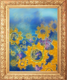
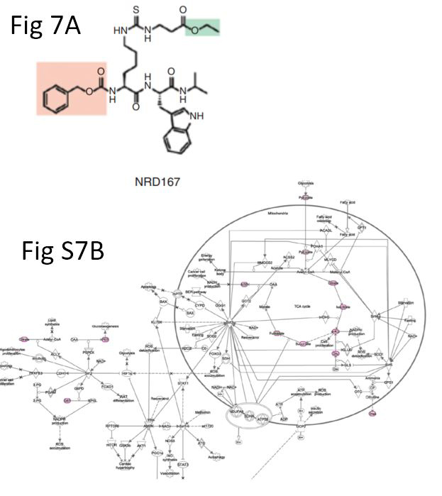
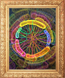
Molecular pathways within our cells operate like machines composed of many gears, in which each gear represents a signaling molecule that interacts with other molecules to drive cell function. The pathways of metabolism are essential for cancer cell survival—if one gear gets jammed, the whole machine will collapse. In this illustration, an inhibitor of the metabolic signaling molecule SIRT5 (Fig 7A) jams the gears of metabolism pathway (Fig S7B), which could disrupt cancer cell growth. Read the full article here.
Untangling skeins of DNA
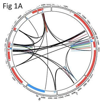
The chromosomes that hold our DNA are packed tightly into each cell’s nucleus, often in close contact with one another. In cancer cells, defects in cell division and DNA repair can cause chromosomes to exchange pieces, a process that researchers visualize using Circos plots like the one shown in Fig 1A. This artistic depiction of a Circos plot hearkens back to the chromosomes in a colorful representation of these DNA exchanges. Read the full article here.
A good catch
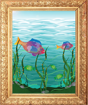
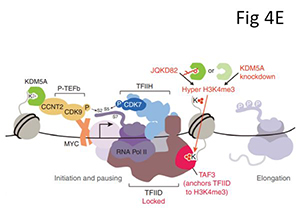
When RNA polymerase II transcribes DNA into protein-coding messages, the polymerase complex (shown in Fig 4E and illustrated as the larger, colorful fish) moves along DNA, taking cues from marks on histone tails. Other enzymes are responsible for regulating these histone marks. If lysine demethylase 5A (green fish) doesn’t remove enough methylation marks (orange dots) from histone tails (seaweed), the polymerase complex becomes stuck and cannot move along DNA, preventing transcription and potentially the growth of cancer cells. Read the full article here.