NCI Collection Offers a Look at “Cancer Close Up”
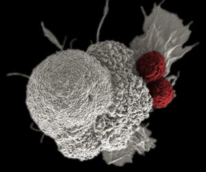
Image by Rita Elena Serda. All images are shared courtesy of the National Cancer Institute.
It looks like a toppled ice cream sundae, with maraschino cherries rolling away from a melting scoop of vanilla.
The reality isn’t even close. The scoop of white is an oral squamous cancer cell, and the red balls are two cytotoxic T cells, attacking the cancer as part of a natural immune response. The image was captured by Rita Elena Serda, PhD, while conducting research on nanomedicine at the Duncan Comprehensive Cancer Center at Baylor College of Medicine. Serda enhanced the microscopy image with Photoshop, using the stark contrast between the red and white to highlight the difference between the cancer cell and the T cells.
The image is part of “Cancer Close Up,” a collection curated by the National Cancer Institute (NCI). The NCI launched Cancer Close Up in 2015 with a collection of images captured by scientists at the NCI Center for Research.
For the 2016 program, the NCI expanded its reach, soliciting images from all 69 NCI-Designated Cancer Centers. They received nearly 100 submissions, and after a selection process that included scientific review by Rick Manrow, PhD, and Rebecca Chasan, PhD, the NCI created an online photo gallery of 85 images from about two dozen cancer centers.
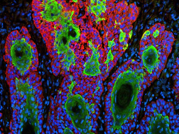
This image, captured by Khalid Mohammad and Theresa Guise at Indiana University’s Simon Cancer Center, illustrates the metastasis of cancer cells to the bone microenvironment from a primary site. Understanding how cancer cells spread to bone and cause bone destruction is important to finding successful treatment, the NCI said.
The images are in the public domain, accessible to anyone via a simple download. They’ve also been on a modest exhibit circuit: The NCI displayed 12 images at the American Association for Cancer Research (AACR) Annual Meeting in April, and 12 different images at the June meeting of the American Society of Clinical Oncologists.
Most of the images originated as potential illustrations for research papers or slide presentations. Over time, it became apparent that many of the images had an abstract, otherworldly beauty. Most pictures were taken with digital cameras attached to fluorescent optical microscopes. Stains were often used to highlight different components of the images, and photo-editing programs were used in some cases.
Cindy Lollar, content manager of NCI Visuals Online, said that in order to be selected for the Cancer Close Up project, images had to have been created by scientists; had to represent important frontiers in cancer research, especially in precision medicine; and had to be visually compelling in color, contrast, and composition.
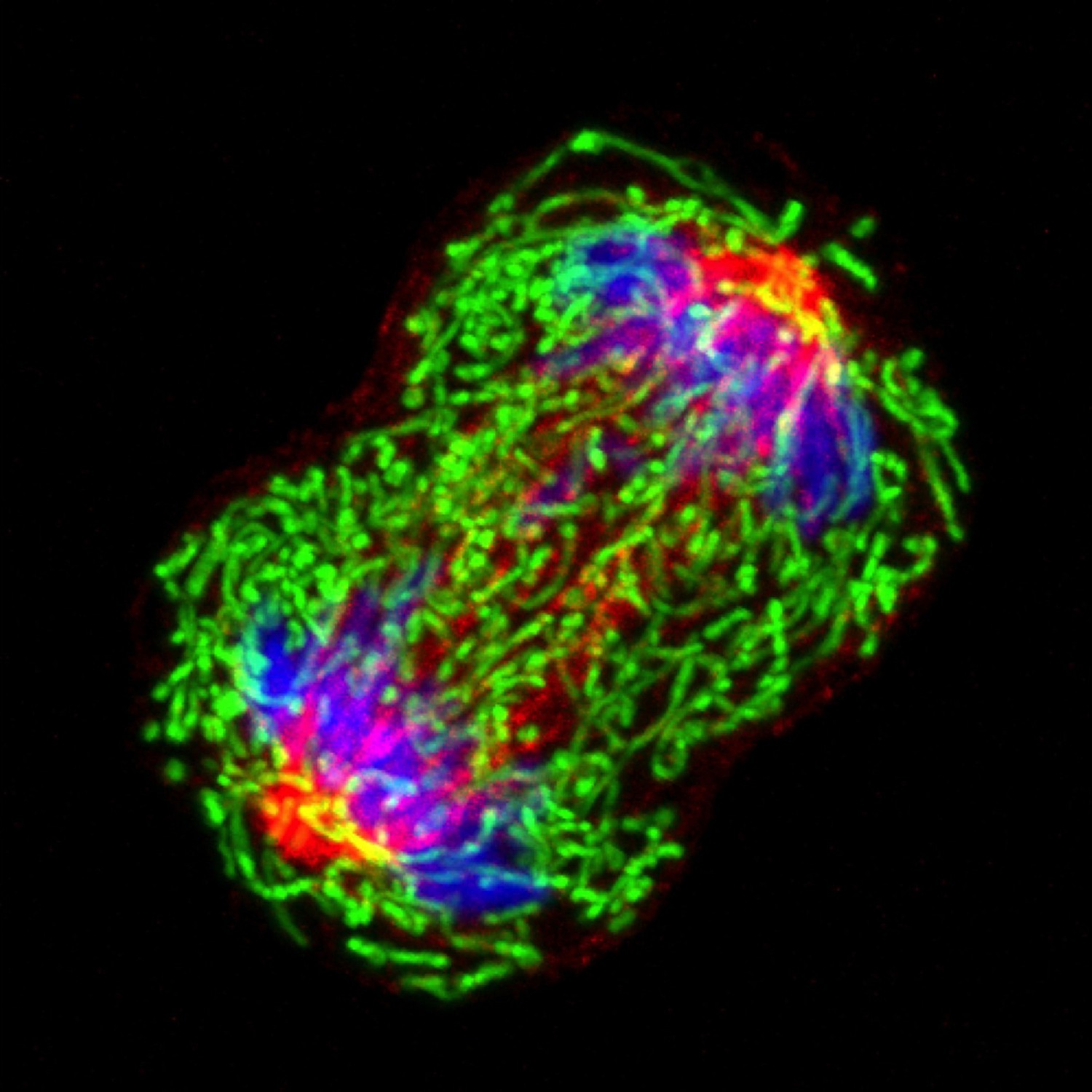
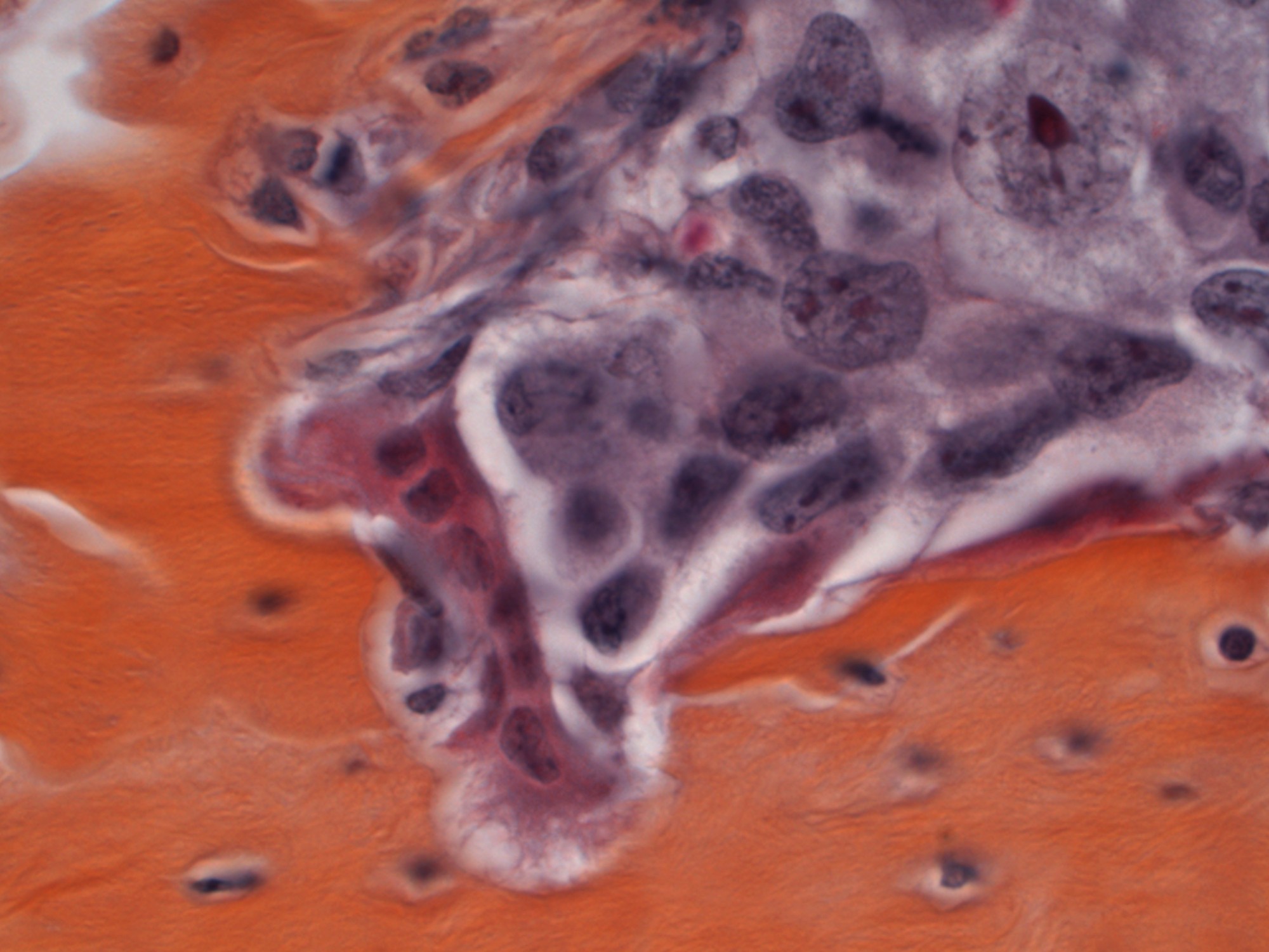
This image, captured by Wei Qian of the University of Pittsburgh Cancer Institute, shows a triple-negative breast cancer cell in metaphase, the second stage of cell division. A better understanding of how mitochondria play roles in tumor cell division may provide new therapeutic targeting strategies, the NCI said.
“In this era of precision medicine, it’s never been more important to understand cancer at the molecular level,” Lollar said. In addition to traveling exhibits like the AACR and ASCO meetings, Lollar anticipates that the images will be used by media outlets and by teachers at all levels, including health educators working with patients and their families.
In addition, Lollar said, “We hope that research advocates and policymakers find the images a helpful way to better understand this important area of cancer research and would feel free to use them in their presentations and publications.”