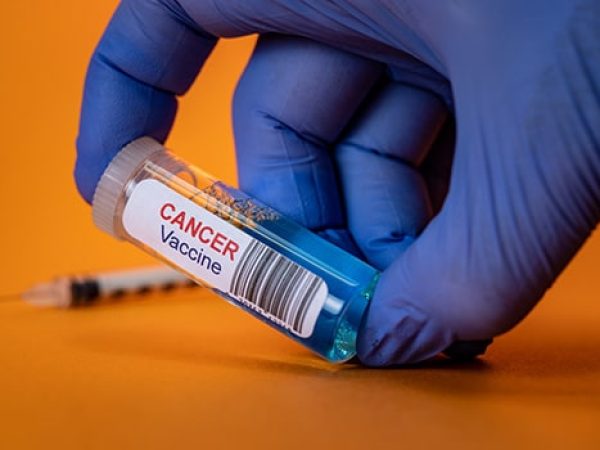
From the Bench, Summer 2025: In Vivo Treatment Spies to Counter Cancer’s Cunning
Cancer is clever and often uses tricks to outsmart both the body’s defenses and treatment strategies. In this summer edition of our “From the Bench” series, we highlight cutting-edge therapeutic technologies that can generate or transform treatments in vivo to counter cancer, including cellular therapies, lactate-sensitive nanoparticle drug delivery, bioink-targeted therapeutics, and click-chemistry protodrugs. Like secret agents that infiltrate behind enemy lines, the four experimental advances featured here involve payloads that only become potent once they are inside the body and receive the right signal. Together, they provide a potential preview of tomorrow’s safer, smarter medicines.
Slipping into Circulation to Generate CAR T Cells in Vivo
Imagine a nanoparticle spy slipping into circulation with classified documents that teach T cells a new way to recognize cancer. A study published in Science explored this concept using targeted lipid nanoparticles (tLNPs) to reprogram T cells into superior cancer-fighting cells. The particles are constructed using a novel ionizable lipid molecule (L829), are conjugated to T‑cell-homing antibodies, and carry mRNA blueprints encoding a CD19‑directed chimeric antigen receptor (CAR). Once they dock and deliver their cargo, the T cells translate the message and display the new receptor, effectively field‑promoting themselves into CAR T cells.

In preclinical testing, the L829 tLNP platform successfully engineered CD8-positive T cells derived from both healthy donors and patients, and demonstrated the ability to control acute lymphoblastic leukemia in humanized mouse models. In healthy monkeys, a similar strategy generated CD20-targeting CAR T cells in vivo that wiped out their B cells and appeared to “reset” their immune system, with few long-lived memory B cells and populations dominated by less experienced B cells.
CAR T-cell therapies have already turned the tide for many patients with otherwise intractable B‑cell leukemias and lymphomas; a single‑shot infusion that installs the same receptors could help spread that success. By eliminating patient‑specific cell collection, viral vectors, conditioning chemotherapy, and centralized manufacturing bottlenecks that restrict access to current CAR T-cell therapies, nanoparticles that can generate treatments in vivo could expand access for more patients with blood cancer. And swapping the mRNA blueprint opens the door to new CAR targets—solid tumor antigens, autoimmune culprits, even fibrotic markers in the heart—that could extend the reach of this platform well beyond blood cancers.
Waiting for Warburg’s Signal: Lactate-responsive Nanoparticle Cancer Therapy
A second nanoparticle spy carries powerful therapeutics under lock and key, trained not to trigger the anticancer device unless they sense the tumor is close. A study published in Cell Reports Medicine demonstrated a strategy that takes advantage of the Warburg effect, a phenomenon where tumors metabolize glucose and secrete lactate, even in the presence of adequate oxygen for aerobic metabolism. This accumulation of lactate is associated with a variety of malignant traits.

In a nod to the two-faced Roman god, investigators built a Janus nanoparticle: on one side, a mesoporous silica cargo face loaded with a cancer therapeutic and sealed by cleavable linkers, and a gold sensor face on the other. In the presence of high levels of lactate, the enzyme lactate oxidase, which is attached to the gold side of the nanoparticle, generates hydrogen peroxide. The peroxide then cuts the molecular cord to the chemotherapy-containing caps and unleashes cancer-killing compounds at the tumor site.
Keying in vivo drug release to a tumor metabolic signature enabled the targeted delivery of chemotherapy and boosted therapeutic effect in mouse models of breast cancer and Ewing sarcoma, compared with free drug and acid‑only controls. Lactate-responsive nanoparticle therapeutic delivery of the STING agonist SR‑717 also shrunk breast cancers more effectively in mice. Moving forward, metabolic gating could help turn a tumor’s lactate plume into a local drug trigger, although hurdles involving enzyme stability, variable lactate levels, and large‑scale manufacturing need to be overcome.
Shaped in Silence: Ultrasound-guided 3D Bioprinting in the Body
Our third covert operative can shift its shape to fit the situation. Instead of surgically implanting a scaffold within patients, what if doctors could inject and navigate a fluid “bioink,” aim an ultrasound wand, and have a customized structure spring up in the exact right place? Even better, what if they could preload structures with anticancer drugs or molecules to help repair or regenerate tissues?

That could be the future, according to a study published in Science that showcases imaging‑guided deep tissue in vivo sound printing (DISP). This method mixes a printable polymer hydrogel with heat-sensitive lipid vesicles that allow the bioink to be tracked and manipulated in the body with ultrasound. Once maneuvered to the site of interest, the bioink is beamed with focused ultrasound that bursts the vesicles and releases the cross-linking agents that allow the structure to materialize.
As a proof-of-concept toward treating bladder cancer, the researchers navigated the ink in vivo to mouse bladders and produced precise patterns on site, suggesting the ability to deliver targeted cancer treatments. Bioinks loaded with the chemotherapy doxorubicin eliminated more cancer cells compared with free drug administration with tumor spheroids in lab studies.
Furthermore, the team demonstrated the technology’s wide range of potential applications by using the same basic approach to create bioinks that were electrically conductive, cell-transporting, or adhesive. No doubt safely translating this technology to humans may prove challenging, but the core idea is compelling: print where you point, and build tailor-made therapeutic outposts inside tumor territory.
Click-chemistry Protodrugs: The Secret Biorthogonal Handshake
Our final schemers work like two spies exchanging information through a secret handshake. The code behind that handshake is click chemistry, and a study published in the AACR journal Clinical Cancer Research highlighted clinical results involving in vivo click chemistry, showing that a chemotherapy protodrug can be guided to and activated at the sites of solid tumors within the human body.

Click chemistry involves a family of ultra‑fast, ultra‑selective chemical reactions where partners “click” together inside living tissue without disturbing anything else. This cancer treatment—SQ3370—utilizes an in vivo click chemistry reaction between tetrazines and strained trans‑cyclooctenes (TCOs). First, doctors inject the tetrazine component, a clickable biopolymer that serves as a pretargeting agent that homes to and concentrates in the tumor. Soon after, they infuse a clickable chemotherapy protodrug, doxorubicin masked by TCO. This payload circulates harmlessly through the body—until it meets and clicks its partner and releases active doxorubicin near the tumor, which could help decrease systemic side effects associated with various treatments.
Doxorubicin, for instance, can cause heart damage in addition to weight loss when administered intravenously due to its systemic spread, even while low levels of the drug accumulate at the cancer site. However, intravenous SQ3370 led to much higher drug concentrations near tumors in mice, and shrunk those tumors while sparing the mice from these side effects. A phase I/II trial escalated the usual human dose to 15 times without hitting a maximum tolerated level, and the drug stayed inactive until reaching the click site, where biopsies showed a surge of CD8-positive T cells, suggesting immune engagement. Because the platform needs only a clickable polymer and a masked payload, it could be retooled for other cytotoxic drugs, immune agonists, or combination cocktails, offering a chemistry‑guided route to safer, localized therapy.