Striking a Nerve in Pancreatic Cancer: Unraveling the Crosstalk Between Tumor Cells and Neurons
One of the hallmarks of pancreatic cancer is a highly complex tumor microenvironment (TME) that plays an essential role in immune evasion, resistance to therapy, and metastasis. A key component of this ecosystem is the relationship between cancer cells and nerve cells.
Perineural invasion, or the ability of cancer cells to invade nerves and travel along them, has been recognized as a feature of pancreatic ductal adenocarcinoma (PDAC) for decades and is an underlining cause of the severe pain experienced by many patients. On the other hand, nerves within the tumor microenvironment grow larger (a process called nerve hypertrophy) and innervate the tumor.
Scientists in the field of cancer neuroscience study the interactions between cancer and the nervous system and how these interactions influence progression of the disease and patient outcomes. In the context of pancreatic cancer, researchers are dissecting the nature of the tumor-nerve connection, which might do much more than just provide a preferential path for tumors to spread locally outside of the pancreas.
The complex biology of this crosstalk was a major theme at the AACR Conference on Pancreatic Cancer: New Frontiers in Biology and Therapeutic Development, held in San Diego on September 25-28, 2026.
What Is the Role of Fibroblasts in the Tumor-nerve Crosstalk?

Eileen Carpenter, MD, PhD, assistant professor of internal medicine at the University of Michigan Medical School, who delivered the Rising Star Keynote, zoomed in on the role of fibroblasts in the interaction between tumor cells and nerves and shared her lab’s efforts to isolate and study the nerves in the human pancreas.
Carpenter’s research on the role of fibroblasts in the tumor-nerve crosstalk stemmed from a molecular profiling study of matched pre- and post-chemotherapy tissue samples from a small cohort of patients with PDAC. A key finding from this study was that a group of genes that control how nerve fibers form and extend was highly expressed in cancer-associated fibroblasts (CAFs) after chemotherapy.
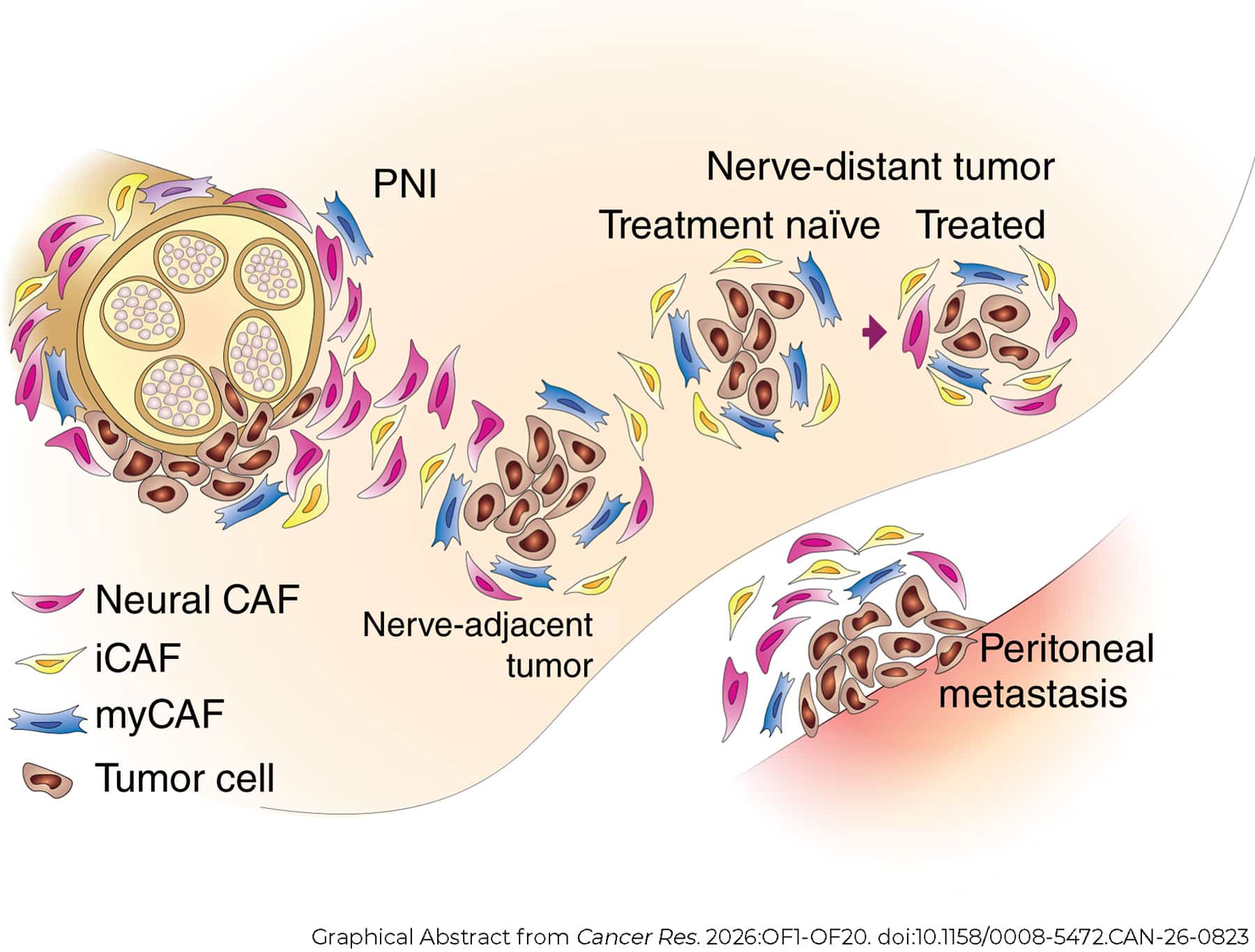
CAFs are a central element in the pancreatic TME. They produce proteins and enzymes that are part of the dense, stiff fibrotic tissue surrounding PDAC tumors and profoundly influence the behavior of cancer cells. CAFs expressing the neural signature, which the researchers dubbed neural CAFs, were observed in areas of perineural invasion and were also more abundant after chemotherapy in areas away from the nerves.
Furthermore, neural CAFs were found to be more prevalent in peritoneal metastasis compared with the primary tumor and other metastatic sites. Notably, the presence of neural CAFs correlated with increased tumor cell proliferation.
These findings, which were recently published in Cancer Research, spurred a number of follow-up questions on the role of nerves in pancreatic cancer.
Exploring the Neural Network of the Human Pancreas
All parts of our body are wired by the peripheral nervous system that carries messages between the brain and spinal cord and the rest of the body, allowing us to sense our environment, move our muscles, and execute involuntary vital functions such as breathing, heartbeat, and digestion.
Carpenter and colleagues analyzed tissue samples from normal human pancreas and pancreatic tumors to study how peripheral innervation differs between healthy and malignant states. They observed that the normal pancreas is highly innervated by different types of neurons: sensory neurons, which detect sensory information in the organs and relay it to the brain and spinal cord; sympathetic neurons, which carry signals from the brain and the spinal cord to the organs to help them respond to stress; and parasympathetic neurons, which carry the information that is needed to control organ function during rest and digestion.
However, nerves in tumor tissues were larger than in the normal pancreas, indicating nerve hypertrophy, and there was an increased proportion of sympathetic neurons than sensory neurons.
Bringing Human Neurons Into the Laboratory
The team then wanted to dissect the function of neurons in PDAC and their interaction with tumor cells and the microenvironment. However, isolating and culturing neurons from tumor tissue poses a unique challenge, because the tumor only houses a portion of the nerve cells, while their bodies, which contain their nuclei, are located outside of the pancreas. They are found clustered together in bundles called ganglia, which serve as communication hubs. Specifically, the sympathetic cell bodies reside in the celiac and mesenteric ganglia, located deep in the abdomen, and the sensory cell bodies reside in the dorsal root ganglia, located along the spine.

Through Michigan’s Gift of Life program that allows nontransplantable tissue from organ donors to be used for research purposes, Carpenter and team obtained human nerve explants from which they isolated celiac ganglia and dorsal root ganglia and grew sympathetic and sensory neuron cells in culture.
While it is very exciting to work with these neural explants, there are several caveats, Carpenter explained. These include variability from explant to explant, the inability to obtain enough cells for large experiments, and the confounding presence of other structures in the explants besides the nerves.
To circumvent these issues, the researchers used a complementary approach and obtained sensory neurons in vitro through differentiation of human induced pluripotent stem cells (iPSCs). Coculture of these “iNeurons” with tumor cells and CAFs led to upregulation of genes encoding inflammatory cytokines, such as IL-8 and CXCL1, and mediators of the NF-kB signaling pathway. “We know that these chemokines play a big role in PDAC and its microenvironment,” Carpenter noted. “Our findings suggest that the neurons could potentially be a source of these inflammatory molecules.”
To continue to dissect perineural invasion in vitro, the team is currently developing iPSC-derived sympathetic neurons to compare them directly with sensory neurons and see whether these two types of neurons play different roles in pancreatic cancer.
Carpenter and colleagues also plan to study the role of glial cells (non-neuronal cells that provide support, nourishment, and protection to neurons) in perineural invasion taking advantage of glial cell lines they were able to grow from the nerve explants.
A Closer Look: Understanding How PDAC Reprograms Neurons
During a plenary session on the desmoplastic and neural microenvironment of pancreatic cancer, Andreas Trumpp, PhD, head of the Stem Cells and Cancer Division of the German Cancer Research Center (DKFZ) and director of the Heidelberg Institute for Stem Cell Technology and Experimental Medicine (HI-STEM), discussed his lab’s research on tumor-neuron interactions in human and mouse models of PDAC.

Trumpp started his talk by emphasizing the growth of the field of cancer neuroscience in the past 10 years, propelled by the publication of fundamental studies demonstrating that disruption of innervation impaired the development of cancer and by the availability of new technologies to study the tumor-nerve crosstalk.
Any tumor that grows within an organ will at some point come into contact not only with fibroblasts, immune cells, and blood vessels, but also with resident neurons, which become components of the TME, Trumpp said. He added that neurons and innervation were recognized as hallmark-enabling components of the TME in the most recent edition of the “Hallmarks of Cancer.”
Since the cell bodies and nuclei of neurons are located far away from the tumor tissue, multiomics analysis of tumor samples does not capture the full molecular characteristics of the neuronal component. To address this, Trumpp’s lab developed a new method for profiling peripheral neurons called Trace-n-Seq. Through this technique, researchers can trace nerves back to their cell bodies, isolate these from the ganglia, and analyze their transcriptomes at the single-cell level.
Using Trace-n-Seq, Trumpp and colleagues compared nerves from the normal pancreas and PDAC. They observed that both sympathetic and sensory neurons from PDAC tissues became reprogrammed by the tumor and expressed genes involved in stemness and encoding axon guidance molecules, which orchestrate the increased growth of the neurons’ branches and promote tumor growth.
Consistent with this finding, studies in different mouse models of pancreatic cancer showed increased axonal sprouting in pancreatic tumor tissue compared with normal pancreas, even though the number of innervating neurons was unchanged.
Furthermore, the researchers observed that some neurons branch out into more than one organ, suggesting that tumor-reprogrammed neurons may also innervate healthy organs, for example the liver, and condition them to establish a premetastatic niche. In other words, these neurons may set the stage for metastatic cells invading from the pancreas to start proliferating in the liver, Trumpp speculated.
Cutting the Line of Communication: Can Disrupting Neural Signals Slow Pancreatic Cancer?
Further dissecting the neuron-tumor cell crosstalk, Trumpp and colleagues showed that PDAC cells cultured in the presence of neurons proliferated more and produced proteins involved in the organization and maturation of synapses, the specialized junctions where electrical or chemical signals pass from one neuron to another cell. Indeed, super-resolution microscopy and electrophysiological studies provided structural and functional evidence for the presence of synaptic connections through which neurons transmit signals to tumor cells.
Next, the team studied the effect of blocking the tumor-nerve communication. In mouse models of PDAC, treatment with a neurotoxin called 6-hydroxydopamine (6-OHDA), which is commonly used to induce sympathetic nerve damage in animal models, slowed down tumor growth.

The researchers then hypothesized that drugs used for first-line treatment of pancreatic cancer, such as nab-paclitaxel (Abraxane), which cause substantial chemotherapy-related neuropathy, may do so by also damaging the sensory neurons within the tumor. If so, Trumpp reasoned, there might be more to these drugs than the ability to kill tumor cells. Indeed, treatment with nab-paclitaxel caused intratumoral neuropathy, specifically targeting the sensory neurons.
Therefore, Trumpp explained, it seemed logical to combine 6-OHDA and nab-paclitaxel to study the effect of denervation of both sympathetic and sensory neurons at the same time. Combination treatment with 6-OHDA and nab-paclitaxel had a synergistic effect on tumor growth, resulting in a much deeper reduction than with either treatment alone.
To translate these findings into the clinic, the team is planning a phase I/II clinical trial called SYNERGY combining frontline chemotherapy, including nab-paclitaxel, with targeting of the sympathetic neurons in patients with locally advanced, unresectable pancreatic cancer. Disruption of the sympathetic neurons can be achieved by delivering high-dose targeted radiation specifically to the celiac plexus through a method called radiosurgery, which is currently used as an effective treatment for pain reduction in patients with pancreatic cancer. One arm of the trial patients will also receive a checkpoint inhibitor, as the team showed that denervation makes PDAC sensitive to immunotherapy in a mouse model. In addition to an increase in overall survival, a secondary goal is to assess whether this triple combination treatment is able to shrink the tumor to a degree that allows surgical resection, Trumpp concluded.
Back in the lab, the team is now expanding the applications of Trace-n-Seq to several other carcinoma models and studying the progression of neuron reprogramming from the primary tumor to metastasis.