Editors’ Picks, May 2026: Insights Into CAR T Therapy, Neuron-Tumor Crosstalk in Pancreatic Cancer, and More
After a rainy Memorial Day weekend here on the East Coast, we are thrilled to see some sunshine this week. Brighten up your day, wherever you are, with the latest edition of Editors’ Picks from the AACR’s 10 peer-reviewed journals. This month, you can read about CD28’s impact on CAR T-cell activity; the evaluation of investigational therapeutics for solid tumors; new insights into the progression of pancreatic, lung, breast, and prostate cancers; factors contributing to disparities in pediatric leukemia outcomes; and more.
Keep reading to learn about this month’s featured studies, and follow the links for the full text of each article, freely available for a limited time.
Journal: Blood Cancer Discovery
Endogenous CD28 Drives the Persistent Activity of CAR T Cells in Myeloma and Lymphoma Models
Chimeric antigen receptor (CAR) T-cell therapy has reshaped the therapeutic landscape for multiple myeloma, yet most patients treated with BCMA-targeted CAR T cells experience disease relapse. Consequently, we sought to determine if inhibition of CD28 survival signaling could increase multiple myeloma sensitivity to CAR T-cell therapy. Contrary to expectations, blockade of CD28 interaction with CD80/86 accelerated tumor regrowth in preclinical multiple myeloma and lymphoma CAR T-cell therapy models. Knockout studies revealed that endogenous CD28 on 4-1BB costimulated CAR T cells prolonged in vivo activity, reprogrammed mitochondrial metabolism to maintain redox balance, and stimulated proliferation and release of tumor model–specific inflammatory cytokines in the tumor microenvironment (TME). Intriguingly, transient CD28 blockade decreased levels of certain TME cytokines without significantly affecting survival of CAR T cell–treated mice. Collectively, these data provide direct evidence that endogenous CD28 signaling modulates CAR T-cell responses in multiple myeloma and lymphoma models.
Significance: This study provides direct evidence that endogenous CD28 on 4-1BB costimulated CAR T cells promotes cytotoxic activity and the production of inflammatory cytokines in the TME. These findings have important implications for ongoing efforts to improve CAR T-cell therapy for the treatment of hematologic malignancies.
The article was featured on the cover of the May issue, which also included a related commentary.
Journal: Cancer Discovery
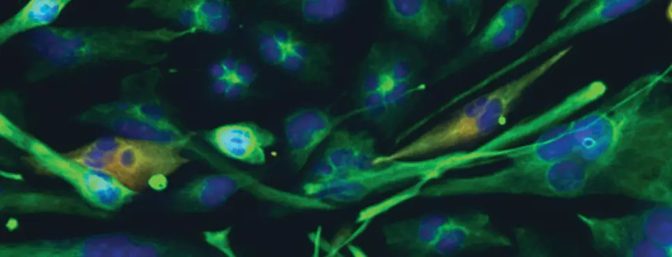
Myofibroblasts Induce Neuroplasticity to Promote Pancreatic Inflammation and Cancer Progression

Pancreatic ductal adenocarcinoma (PDAC) co-opts the peripheral nervous system through nerve hypertrophy, axonogenesis, and perineural invasion, and these processes correlate with patient morbidity and mortality. Prior work has shown that autonomic nerves directly modulate neoplastic cells in PDAC, but whether cancer-associated fibroblasts (CAF) participate in neural remodeling is unknown. Using thick tissue sections, we identified dense neo-innervation near myofibroblastic CAFs (myCAF) in preinvasive pancreatic intraepithelial neoplasms. Mechanistically, TGFβ produced during inflammation and neoplasia triggers myofibroblast formation, and myCAFs produce axon guidance molecules that recruit sympathetic nerves. Norepinephrine released by sympathetic nerves activates myofibroblast cultures in vitro, and sympathetic nerve depletion impairs stromal activation and PDAC growth in vivo. A chemogenetic model confirmed that fibroblast-specific α1-adrenergic signaling exacerbated pancreatic inflammation and neoplasia. Therefore, beyond direct epithelial effects, sympathetic nerves promote pancreatitis and PDAC by co-opting myofibroblasts and myCAFs as disease amplifiers, highlighting CAF subtype–specific stromal interactions as putative therapeutic targets.
Significance: Pathology-associated myofibroblasts orchestrate bidirectional cross-talk with sympathetic neurons, secreting axon guidance molecules to promote nerve infiltration in inflamed and neoplastic pancreatic tissues. Specifically, α1-adrenoreceptor activation in fibroblasts acts as a molecular switch that amplifies pancreatitis severity and accelerates tumor growth, revealing new paracrine and juxtacrine interactions for further therapeutic development.
The article was featured on the cover of the May issue, which also included a related commentary.
Journal: Cancer Epidemiology, Biomarkers & Prevention
Background: Pediatric acute lymphoblastic leukemia (ALL) is the most common cancer in children, and its incidence and outcomes vary by race and ethnicity.
Methods: The Reducing Ethnic Disparities in Acute Leukemia (REDIAL) Consortium has assembled a cohort of patients <25 years at diagnosis to examine genetic and neighborhood-level factors influencing ALL. As the cohort is ongoing, for this report, we examined sociodemographic and cytogenetic factors that might be associated with ALL outcome disparities.
Results: Among 2,512 patients diagnosed between 2005 and 2020, we observed distinct patterns of genetic ancestry and residential characteristics across self-reported race and ethnicity. Latino children, who comprised 55.3% of the cohort, had a mean Amerindigenous ancestry proportion of 50.9%, whereas non-Latino White children exhibited a predominantly European ancestry (mean = 78%). Neighborhood-level analysis revealed significant socioeconomic and geographic differences by race/ethnicity, with Latino children more frequently residing in Latino enclaves or economically disadvantaged neighborhoods. Cytogenetic profiling of 551 patients showed that favorable subtypes (double trisomies and ETV6::RUNX1) predominated overall, but ETV6::RUNX1 was significantly less common in Latino than in non-Latino White children; conversely, CRLF2 overexpression and immunoglobulin heavy chain (IGH) rearrangements were more frequent among Latino children.
Conclusions: This first REDIAL cohort analysis reveals how genetic ancestry and neighborhood-level factors jointly shape the distribution of ALL cytogenetic subtypes, advancing our understanding of contributors to disparities in ALL outcomes.
Impact: This analysis offers insights into factors underlying disparities in ALL outcomes that may be targeted for mitigation, ultimately guiding more equitable approaches to risk stratification and intervention.
Journal: Cancer Immunology Research
IL10R Inhibition Induces Neutrophil Tumoricidal Activity
Although the role of neutrophils in modulating antitumor T-cell responses has been extensively studied, their direct effects on tumor cells remain less well understood. In this study, we investigated whether neutrophils have the capacity to directly kill tumor cells independently of T cells. We found that anti-CD40–based therapy, when combined with IL10 receptor blockade, initiates a Batf3-dependent pathway in which IL12 and IFNγ secretion results in oncolytic neutrophil activity. Using a combination of microscopy, single-cell, and functional assays, we observed that killing of tumor cells by neutrophils is dependent on physical contact and degranulation. This degranulation-mediated killing is associated with an atypical dynamic invasive neutrophil phenotype. In line with our preclinical findings, our phase I trial of anti-CD40 shows that circulating IL12, IFNγ, and IL10 increase in response to anti-CD40, whereas our phase Ib/2 PRINCE study shows that lower circulating IL10 is associated with favorable overall survival (OS) specifically among anti-CD40–treated patients. Finally, we found that neutrophil expansion with granulocyte colony-stimulating factor is associated with improved OS, specifically in patients treated with anti-CD40, suggesting that this pathway may be amenable to therapeutic intervention in patients with advanced cancer.
Journal: Cancer Prevention Research
Bench-to-Bedside Evaluation of Sulforaphane/BroccoMax on Fatty Acid Synthesis in Prostate Cancer
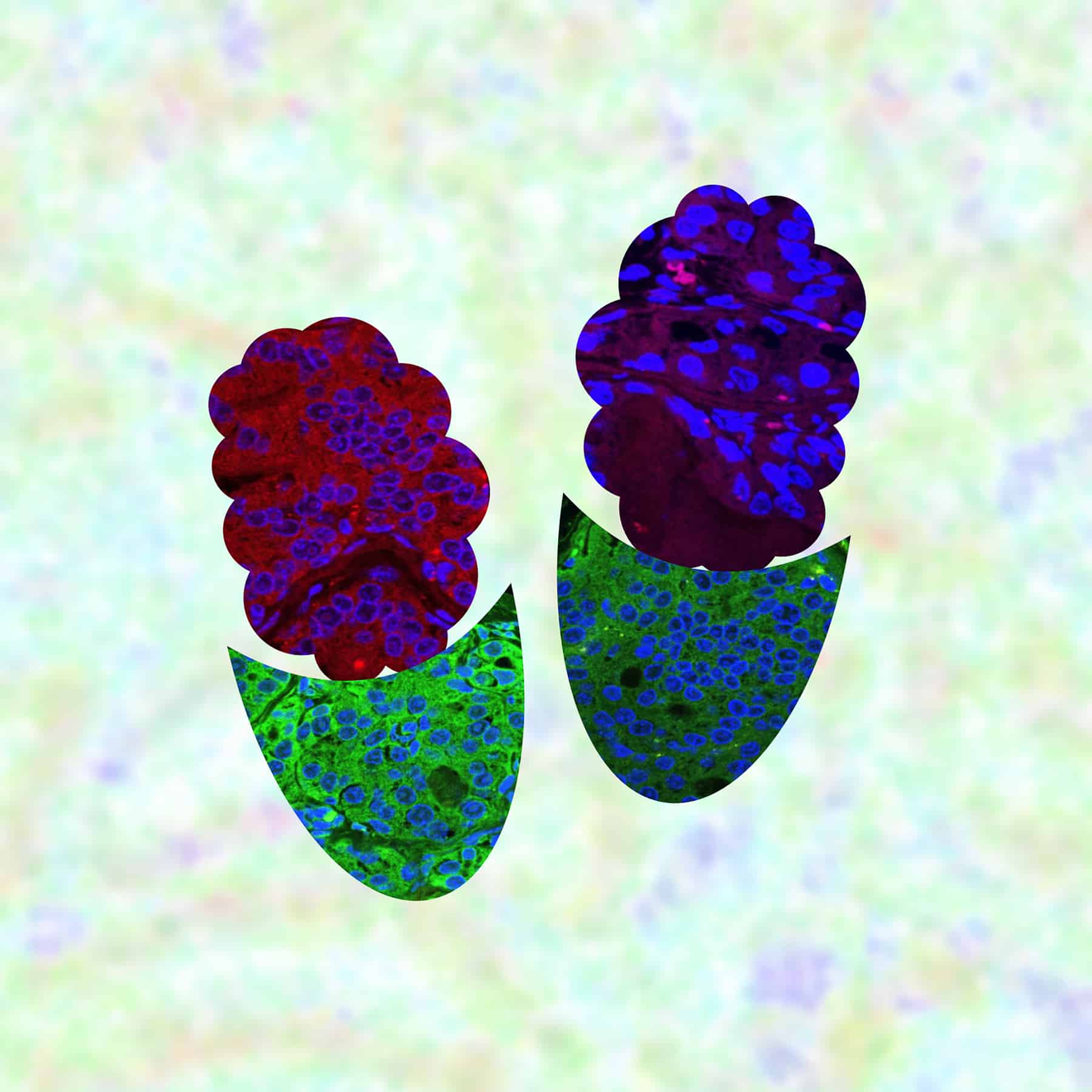
The fatty acid synthesis pathway is a valid target for the prevention of prostate cancer. However, a clinical-grade inhibitor of fatty acid synthesis is still lacking. This bench-to-bedside study was undertaken to determine the feasibility of fatty acid synthesis inhibition using broccoli constituent sulforaphane (SFN) and its clinical-grade formulation BroccoMax (BMAX). Oral administration of SFN to Hi-Myc mice resulted in the inhibition of prostate adenocarcinoma burden by about 61% that was accompanied by a significant decrease in prostate tumor levels of c-Myc and proliferating cell nuclear antigen proteins and increased apoptosis. Expression of acetyl-CoA carboxylase 1 (ACC1) and fatty acid synthase (FASN) was lower by about 46% and 31%, respectively, in the prostate tumor of SFN-treated mice when compared with that of control mice (P < 0.001). Plasma levels of total free fatty acids (TFFA), cholesterol, and total phospholipids were decreased significantly following SFN treatment. In a double-blind clinical trial, patients with histologically confirmed prostate cancer were randomized to the BMAX (n = 19) or placebo (PBO) group (n = 22). Patients were treated with four capsules of BMAX or four capsules of matching PBO orally two times daily after breakfast and dinner for 4 weeks. Prostate tumor expression of c-Myc, ACC1, FASN, and Ki-67 proteins was significantly lower in the BMAX arm when compared with the PBO group. However, the serum or prostate tumor level of acetyl-CoA or TFFA was not decreased by BMAX treatment. A longer duration treatment with BMAX in patients with early-stage prostate cancer may be necessary to lower the circulating or prostate tumor level of TFFA.
Prevention Relevance: Fatty acid synthesis is a valid target for the prevention of human prostate cancer. In this study, we determined the feasibility of fatty acid synthesis inhibition using SFN in a mouse model and BMAX, its clinical-grade formulation, in prostatectomy patients.
This article was featured on the cover of the May issue.
Journal: Cancer Research (May 1 issue)
VGF-Mediated Mitochondrial Remodeling Fuels Lung Adenocarcinoma Brain Metastasis
Lung adenocarcinoma cells exhibit a marked propensity for brain metastasis, in which they face unique metabolic challenges imposed by the microenvironment. The mechanisms that enable lung adenocarcinoma cells to adapt to these constraints represent potential therapeutic targets. In this study, we identified the neuropeptide VGF as a clinically relevant driver of brain metastatic progression. VGF expression was markedly upregulated in lung adenocarcinoma brain metastases, and elevated VGF expression was associated with an increased risk of brain metastasis and poor survival outcomes. Moreover, brain-derived signals induced VGF expression in lung adenocarcinoma cells, promoting cell survival and proliferation through enhanced mitochondrial oxidative phosphorylation and ATP production. Mechanistically, the N-terminal domain of VGF—a nonclassically secreted peptide—interacted with the hydrolase domain of ABHD12B to suppress cardiolipin degradation, leading to increased cardiolipin levels, stabilization of mitochondrial membranes, and a shift toward mitochondrial fusion over excessive fission. These changes helped meet the heightened energetic demands of metastatic cells in the brain. Genetic deletion of the VGF N-terminal domain disrupted mitochondrial fusion, impaired oxidative metabolism, and significantly reduced brain colonization in mouse models of brain metastasis. Together, these findings uncover a role for VGF and establish the VGF–ABHD12B–cardiolipin axis as a critical mechanism underlying metabolic adaptation in lung adenocarcinoma brain metastases, highlighting the potential of this pathway as a therapeutic target for intervention.
Significance: The N-terminal domain of VGF is required for sustaining mitochondrial fusion and oxidative phosphorylation to support metabolic adaptation in lung cancer brain metastases, underscoring a potentially targetable mechanism to impair brain colonization.
Journal: Cancer Research (May 15 issue)
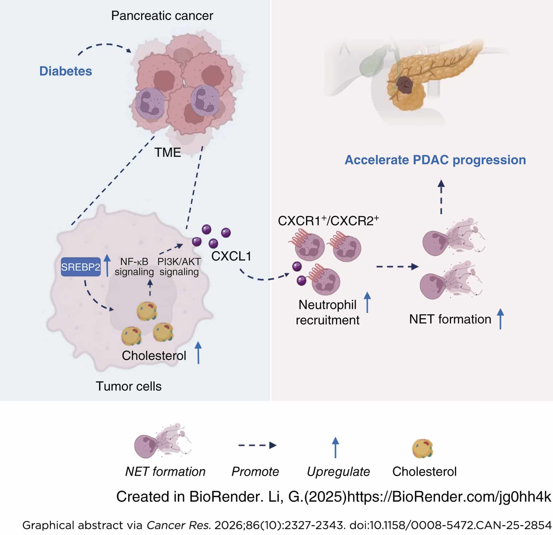
Patients with pancreatic ductal adenocarcinoma (PDAC) with diabetes mellitus (DM) exhibit poor clinical outcomes. Metabolic reprogramming of both cancer cells and immune compartments plays a crucial role in shaping the antitumor immune response in PDAC. DM-induced metabolic alteration may disrupt the intricate cross-talk between immune cells and tumor-associated immune factors, profoundly influencing PDAC progression. In this study, we performed an integrated, spatially resolved multiomics study to investigate DM-associated, cell-specific metabolic remodeling within the PDAC tumor microenvironment. DM influenced interactions between tumor cells and immune cells, which accelerated PDAC growth in both humans and mice. Patients with PDAC with DM exhibited higher tumor stage, poorer differentiation, and worse outcomes. Spatial metabolic and transcriptional profiling revealed that SREBP2-dependent cholesterol biosynthesis exacerbated PDAC progression. Increased cholesterol biosynthesis promoted neutrophil recruitment and accelerated the formation of neutrophil extracellular traps (NET) by stimulating the CXCL1/CXCR1/CXCR2 signaling axis, ultimately promoting PDAC growth. Inhibition of SREBP2, pharmacologic blockade of CXCL1, or perturbation of NETs markedly reduced PDAC growth in diabetic mouse models. Together, these multiomics analyses and follow-up mechanistic studies constitute an integrated approach that elucidates a metabolic mechanism by which diabetes promotes PDAC development by remodeling the tumor-immune microenvironment and highlights a potential therapeutic strategy for PDAC with DM.
Significance: Diabetes induces metabolic reprogramming that promotes neutrophil recruitment and neutrophil extracellular trap formation to drive pancreatic cancer progression, providing a targetable metabolism-immune axis to improve the outcomes of diabetic pancreatic cancer patients.
Journal: Clinical Cancer Research (May 1 issue)
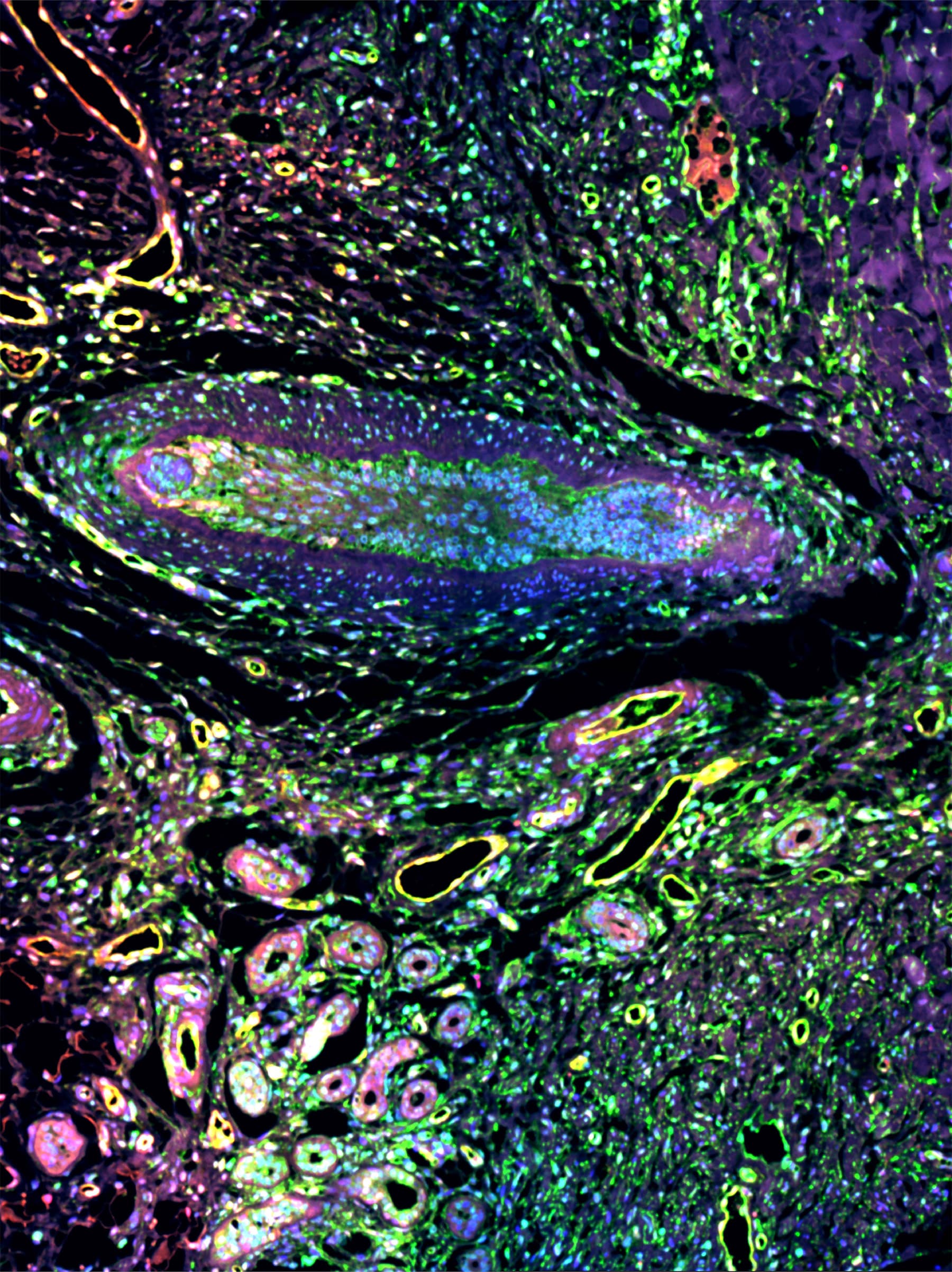
Purpose: This study investigates a short-course, intensified regimen combining apalutamide, abiraterone acetate, and prednisone (AAP) and stereotactic body radiotherapy (SBRT) to reduce treatment burden and improve disease control in a very high-risk (VHR) population inadequately represented in prior trials.
Patients and Methods: This multi-institutional, single-arm, phase 2 trial enrolled patients with VHR localized prostate cancer, defined according to the National Comprehensive Cancer Network as histologically confirmed adenocarcinoma with ≥2 high-risk features: Gleason score 8 to 10, prostate-specific antigen (PSA) ≥20, clinical or radiographic ≥T3, or >4 cores containing Gleason score 8 disease. Patients received 6 months of apalutamide, abiraterone acetate, and leuprolide plus prostate/seminal vesicle–directed ultra-fractionated SBRT. The primary endpoint was 3-year biochemical recurrence (BCR) rate by Phoenix criteria, with a prespecified superiority threshold of <10%. Secondary endpoints included PSA ≥0.2 ng/mL, metastasis-free survival (MFS), and time to testosterone recovery >150 ng/dL.
Results: Between August 2016 and December 2022, 63 patients were treated. At 3 years, the Phoenix-defined BCR rate was 19%. BCR-free survival was 84.2% [95% confidence interval (CI), 75.6–93.7] with a median follow-up of 41 months (34–43). The 3-year MFS was 93.6% (95% CI, 87.8%–99.8%), with no deaths observed. The median time to testosterone recovery >150 ng/dL was 6 months (range, 3–24 months). No new safety signals emerged, and the only significant quality-of-life (QOL) decline was in the EPIC sexual subdomain at 12 months.
Conclusions: Treatment intensification with apalutamide, AAP, androgen deprivation therapy, and SBRT was well-tolerated with limited impact on QOL. Although BCR rates exceed the superiority threshold, outcomes aligned with historic benchmarks, supporting further evaluation of the regimen in prospective trials.
Journal: Clinical Cancer Research (May 15 issue)
Purpose: Plexiform neurofibromas (PN) present a significant clinical challenge, with a notable unmet need for effective and well-tolerated therapies, particularly for the adult population.
Patients and Methods: A single-arm, open-label, phase IIa trial of tunlametinib was conducted to assess its efficacy and safety in adults with inoperable, radiologically measurable PN. Patients received tunlametinib at a dose of 9 mg twice daily, following a continuous 21-day cycle regimen. The primary endpoint was the confirmed objective response rate (ORR), and secondary endpoints included disease control rate, duration of response, progression-free survival, and improvements in patient-reported outcomes (PRO) compared with baseline. Exploratory analyses integrating clinical variables and single-cell transcriptomic profiles were performed to explore response heterogeneity.
Results: Of 15 adults (10 males and five females; median age, 27 years; range, 18–45), eight patients achieved a confirmed partial response, with an ORR of 53.3% (95% confidence interval, 26.69–78.73) and a median neurofibroma volume reduction of −23.5% (range, 1.6% to −49.1%). Significant improvements in multiple PRO domains were observed, including Patients’ Global Impression of Change and the Plexiform Neurofibromas Quality of Life scale. Regarding safety, all patients experienced at least one treatment-related adverse event, the majority of which were grade 1 or 2. Single-cell RNA sequencing profiling of pretreatment biopsies from one rapid responder and two gradual responders revealed substantial microenvironmental heterogeneity.
Conclusions: Tunlametinib demonstrated promising efficacy and an acceptable safety profile in adults with PN, broadly aligning with the prior PN trials, including selumetinib and mirdametinib.
This article was featured on the cover of the May 15 issue.
Journal: Molecular Cancer Research
As cancer cells evade therapeutic pressure and adopt alternate lineage identities not commonly observed in the tissue of origin, they likely adopt alternative metabolic programs to support their evolving demands. Targeting these alternative metabolic programs in distinct molecular subtypes of aggressive prostate cancer may lead to new therapeutic approaches to combat treatment resistance. We identify the poorly studied metabolic enzyme oxoglutarate dehydrogenase-like (OGDHL), named for its structural similarity to the tricarboxylic acid (TCA) cycle enzyme oxoglutarate dehydrogenase, as an unexpected regulator of tumor growth, treatment-induced lineage plasticity, and DNA damage in prostate cancer. Although OGDHL has been described as a tumor suppressor in various cancers, we find that its loss impairs prostate cancer cell proliferation and tumor formation. Loss of OGDHL reduces nucleotide synthesis, induces accumulation of the DNA damage response marker γH2AX, and alters androgen receptor inhibition–induced plasticity. Our data suggest that OGDHL has minimal impact on TCA cycle activity and that mitochondrial localization is not required for its regulation of nucleotide metabolism. Finally, we demonstrate that OGDHL expression is tightly correlated with neuroendocrine (NE) differentiation in clinical prostate cancer and that knockdown of OGDHL impairs the growth of cell line models of NE prostate cancer. These findings underscore the importance of investigating poorly characterized metabolic genes as potential regulators of distinct molecular subtypes of aggressive cancer.
Implications: OGDHL emerged as an unexpected metabolic dependency associated with lineage plasticity and NE differentiation, implicating poorly studied metabolic enzymes as potential targets for treatment-resistant prostate cancer.
Journal: Molecular Cancer Therapeutics
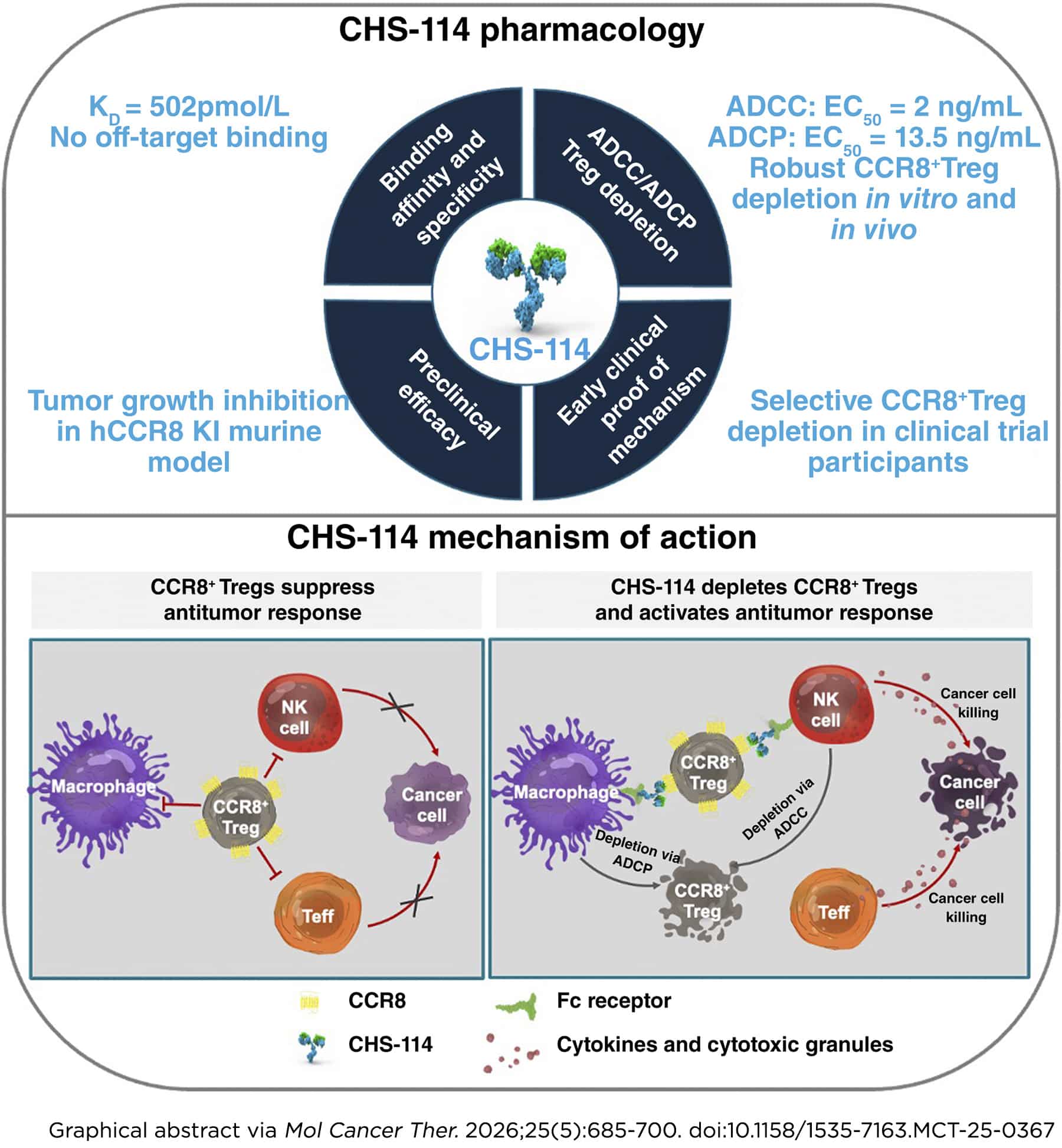
Intratumoral T regulatory cells (Treg) promote an immunosuppressive tumor microenvironment and are frequently associated with a lack of response to immunotherapy. Selective targeting of intratumoral Tregs while sparing broader Tregs and effector T-cell populations is an attractive strategy to enhance antitumor immune responses. C-C motif chemokine receptor 8 (CCR8) is a G protein–coupled receptor that is predominantly upregulated on tumor-resident Tregs in a range of human solid tumors, making it a promising target for their selective depletion. In preclinical studies using mouse tumor models, anti-mouse CCR8 antibody treatment resulted in depletion of CCR8+ intratumoral Tregs, significant antitumor activity, and enhanced survival in combination with anti–PD-1. CHS-114 is a highly selective, afucosylated human anti-CCR8 monoclonal antibody that is being developed as a cancer immunotherapy. CHS-114 selectively binds human CCR8 and potently kills CCR8 expressing cells by inducing antibody-dependent cellular cytotoxicity and antibody-dependent cellular phagocytosis. Ex vivo studies evaluating human dissociated tumor cells demonstrated the selectivity of CHS-114 in depleting intratumoral Tregs while sparing CCR8-negative Tregs and effector T cells. Treatment of tumor-bearing human CCR8 knock-in (huCCR8KI) mice with CHS-114 resulted in significant tumor growth inhibition (62.6%) accompanied by remodeling of the tumor-immune microenvironment and enhanced differentiation of a subset of cytotoxic CD8+ T cells. Based on the promising preclinical data, we are evaluating CHS-114 in clinical trials as an investigational agent for the treatment of solid tumors with and without the anti–PD-1 antibody toripalimab (NCT05635643 and NCT06657144).
Journal: Cancer Research Communications
When tumor cells colonize distant organs during metastasis, they interact extensively with surrounding cells. These interactions often change the behavior of surrounding cell populations which collectively induce a protumor microenvironment that permits tumor cell outgrowth into overt, clinically detectable metastatic disease. The lung is one of the most common sites of breast cancer metastasis. A chronic wound repair–related phenotype developed within the lung microenvironment during metastatic outgrowth in immunocompetent preclinical mouse models of breast cancer. This phenotype was characterized by an increased number and activation of lung type II alveolar epithelial (AT2) cells surrounding growing metastases. Metastatic outgrowth significantly changed AT2 gene expression, resulting in a modified secretome. AT2-derived secreted factors also promoted triple-negative breast cancer growth. AT2-secreted factors are regulated by the cyclic adenosine monophosphate response element-binding protein (CREB). Targeting CREB signaling with the phosphodiesterase 4 (PDE4) inhibitor roflumilast reduced AT2 breast cancer reciprocal interactions in vitro and metastatic outgrowth in vivo. Finally, AT2 cells adjacent to metastases in lungs from patients with metastatic breast cancer expressed higher PDE4B compared with AT2 cells in normal lungs.
Significance: Alveolar epithelial cells are the most common cell type in the lung. Our studies demonstrate the potential for targeting metastasis-associated wound repair and lung epithelial cell activation during metastatic outgrowth with FDA-approved PDE4 inhibitors. This strategy may be an effective way to treat and manage progression of established metastatic breast cancer within the lung.