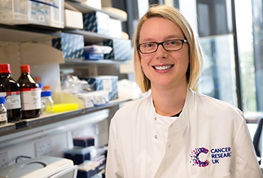
From Stars to Cancer: The Journey of Sarah Bohndiek
By Richard Lobb
AACR Communications
Sarah Bohndiek, PhD, grew up in southeast London, not far from the Royal Observatory Greenwich and the prime meridian, the line of zero degrees longitude that divides east and west. Inspired by the observatory, she wanted at first to become an astrophysicist. She eventually chose a more terrestrial concentration — one in radiation physics. Today she straddles another imaginary dividing line, the one between physics and biology, and uses them both to investigate ways to detect cancer.

As an undergraduate in natural sciences at the University of Cambridge, she took a course in materials science and was introduced to biomaterials. The idea of making artificial blood vessels and joints intrigued her.
“I found it really interesting how someone could use their knowledge from physical sciences to impact everyday lives and really make groundbreaking changes to individual patients,” she recalls, “so that set me thinking about medical physics.”
As a star-struck child, she had also lost her grandmother to breast cancer, and that stayed with her, too.
“One of the areas I thought might be really interesting to work in the medical physics perspective was in cancer, and that was the first place I started with an internship, and I’ve continued ever since developing my career in applying physics to cancer,” she says.
To expand her research horizons beyond the UK, she headed off as a postdoc to Stanford University and the lab of the late Sanjiv Sam Gambhir, MD, PhD, a physicist who became a physician and a leader in molecular imaging and early cancer detection. To support her work in molecular imaging of ovarian cancer, she applied for and received the 2012 AACR-Amgen, Inc. Fellowship in Clinical/Translational Cancer Research.
She appreciated the fact that the AACR-Amgen grant was open to U.S. citizens and non-citizens alike and says the grant was critical from both the scientific and career-building points of view.
“The AACR grant was really pivotal for me at that point in my career, because it gave me the independence to pursue some new lines of research inquiry which I otherwise wouldn’t have necessarily been able to do, but it also gave me the ability to build a network through AACR, going to the AACR meetings and so on. It also gave me a demonstrable track record of securing funding, which is pivotal when you’re trying to apply for faculty positions.”
A faculty position indeed came next, when she joined the Cavendish Laboratory at Cambridge in 2013. Bohndiek has attained the rank of professor at Cambridge and holds a joint appointment in the Department of Physics and the Cancer Research UK Cambridge Institute, with about twenty researchers in her lab. She also leads an international consortium on standardization of photoacoustic imaging which is working to accelerate clinical translation of the technology.
Her lab explores, among other topics, ways to use imaging techniques to understand the microenvironment surrounding a tumor. Of particular interest is the vasculature – the blood vessels that spread through the microenvironment to support the growth of the cancer.
“There’s this phenomenon around an angiogenic switch in which the tumor exceeds the local capacity of oxygen supply, based on its demand, and then tries to recruit new blood vessels into the tumor,” Bohndiek explains. The new blood vessels weave through an emerging, chaotic landscape of nerve paths and muscle fibers that change the composition of the tissue in unpredictable and mystifying ways.
Such vascular alterations can be studied in the gastrointestinal tract using an endoscope, an imaging system in which a light source and a camera are passed inside the patient to relay out images. Bohndiek has modified these endoscopes to enable them to examine a full spectrum of colors rather than just the white light reflectance normally used.
“Many different molecules in our bodies absorb light in different ways,” she says. “In spectral imaging, we measure how light of a range of different colors interacts in the tissue, and by doing so, we can infer what changes in molecular composition have occurred.”
In a study of Barrett’s esophagus (a risk factor for esophageal cancer) published in AACR’s Cancer Research, Bohndiek’s team achieved a 9-fold contrast enhancement for subtle changes that could signal cancer by using a spectrum of colors instead of the standard-of-care white light.
“Hemoglobin in our blood is a strong absorber, and it absorbs the light differently depending on whether it’s well oxygenated or not,” she says. “The changes that we observed in the interaction of light with the tissue showed us that actually there are characteristic changes that suggest the early development of cancer.” The souped-up endoscope could also be used to check the effectiveness of treatment, she says.
Melding physics and biology hasn’t been easy, she admits. She had to learn the language of life sciences and understand the different ways biologists and physicists look at problems. Biologists are always trying to test a hypothesis, she notes.
“In measurement sciences, we aren’t always forming a hypothesis when we go out to do an experiment,” she says. “Sometimes the experiment, in and of itself, is the research. We are working out if it is possible to measure a given phenomenon, and what is the best way to do so.”
Even the process of applying for the AACR-Amgen fellowship helped her bridge the gap between disciplines, she says.
“It was really an opportunity to immerse myself in a completely new field of research. I was looking to write a proposal in an area that I’d never worked in before using model systems that I’d never used before, and methodologies that were completely new to me in many different ways,” she says
“Being able to get that support and that funding from AACR allowed me to completely expand my skill set as a physicist. I was in the lab doing animal handling, developing tumor models, taking blood samples, doing immunohistochemistry — completely on the other side of the fence from my prior experience,” she says. “In addition to that support, there was the kind of the recognition by AACR that made me, as a physicist, feel that I had found a home in the cancer research community.”
##
Work mentioned in the article:
Photoacoustic Tomography Detects Early Vessel Regression and Normalization During Ovarian Tumor Response to the Antiangiogenic Therapy Trebananib
Sarah E. Bohndiek, Laura S. Sasportas, Steven Machtaler, Jesse V. Jokerst, Sharon Hori, and Sanjiv S. Gambhir
J Nucl Med 2015; 56:1942–1947
DOI: 10.2967/jnumed.115.160002
Spectral Endoscopy Enhances Contrast for Neoplasia in Surveillance of Barrett’s Esophagus
Dale J. Waterhouse, Wladyslaw Januszewicz, Sharib Ali, Rebecca C. Fitzgerald, Massimiliano di Pietro, and Sarah E. Bohndiek
Cancer Res 2021;81:3415–25
DOI: 10.1158/0008-5472.CAN-21-0474
The IPASC data format: A consensus data format for photoacoustic imaging.
Gröhl J, Hacker L, Cox BT, Dreher KK, Morscher S, Rakotondrainibe A, Varray F, Yip LCM, Vogt WC, Bohndiek SE, Members of the International Photoacoustic Standardisation Consortium (IPASC)
Photoacoustics, 26 Feb 2022, 26:100339
DOI: 10.1016/j.pacs.2022.100339